Those Wonderful Legs
Margaret Thomas NDB
Margaret Thomas NDB
The inner surface of the basitarsus have hairy brushes. In the first pair of legs they are used to clean dust, pollen and other substances from the head. These legs are then placed between the midlegs and drawn forward, depositing the pollen on the mid leg brushes. The mid legs clean the thorax and transfer pollen to the hind legs.
The first and third legs have specific adaptations. The first leg has an adaptation whereby the fibula is used as an antenna cleaner. Observe a bee cleaning her antennae, placing it in the groove of the tarsus and flexing the tibia-tarsal joint, so closing the trap with the fibula. Then the antenna is drawn through the hole and there it is cleaned, all dust and pollen removed.
Photo 4 First Pair of Legs Adaptions
Scientific Beekeeping
Scientific Beekeeping
info@scientificbeekeeping.co.uk
Human legs are from the top down: Pelvis to which the femur is attached in the hip joint made of a socket, the acetabulum, and the ball (the head of the femur) which has a protuberance called the trochanter to which some of the hip muscles are attached. Then the long bone the femur, joined at the knee joint to the tibia and fibula and at the ankle to the bones of the foot the tarsi.
The bee has bones in the legs named similarly. Starting with the coxa, the trochanter, the femur, the tibia, a tiny fibula and then the five segments of tarsi divided into the large basitarsus and six tarsomeres (the final one being the foot, the surface of which is covered by the tarsal gland epithelium ).
Image 1 Anatomy of a Typical Honey Bee Leg
The end structures are important for walking - the tarsal claws and pads. These are used in walking and also manipulating wax. The suction creating pad, the arolium, allows wakling on horizontal and vertical surfaces.
Image 2
Image 2
Photo 3 Lateral View of Foot
To get the pollen back to the back legs each mid leg is drawn through the hind legs and the brushes collect the pollen onto the hind leg brushes.
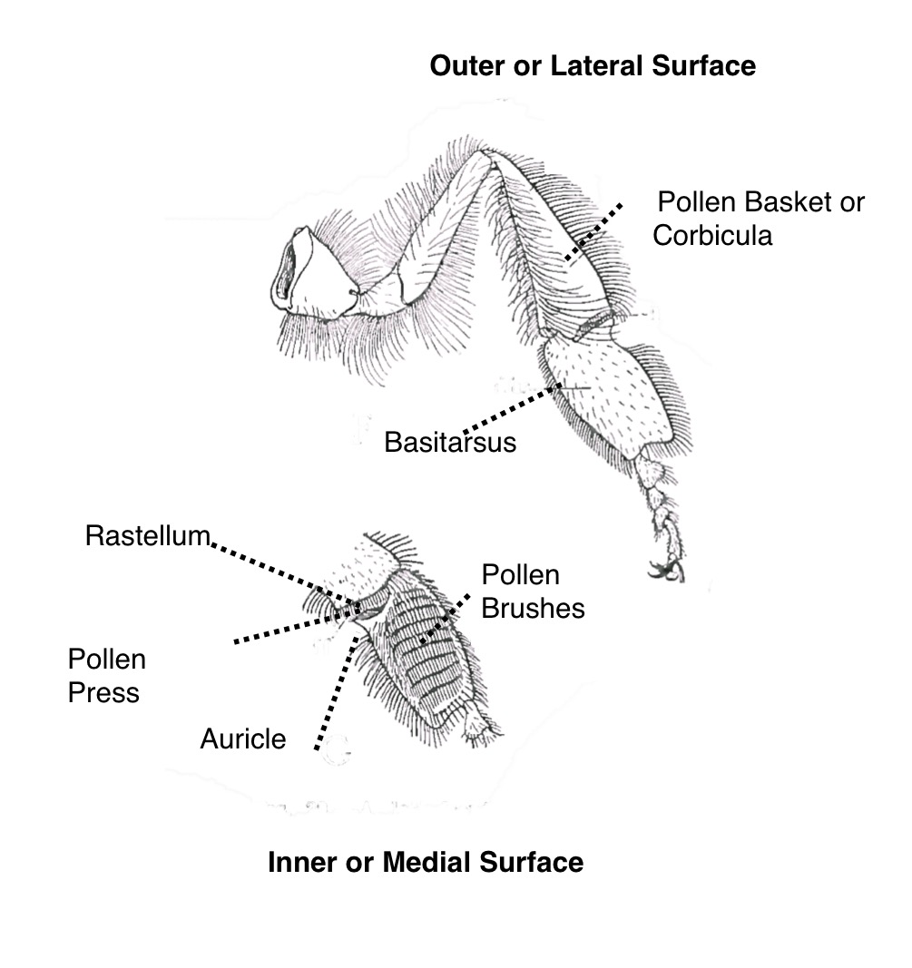
The hind leg is specifically designed to collect pollen and propolis. The parts that are modified are the basitarsus and tibia. The tibia is concave surrounded by long hairs to make a basket called the corbicula. The bottom of the tibia is flattened into a pollen press. When the tibio-tarsal joint is closed the pollen is pushed up into the corbicula. To get the pollen into the pollen press, stiff hairs (rastellum) on the inner surface of the tibia scrape down the stiff bristles of the basitarsus of the opposite leg. Once pushed into the corbicula a central hair acts as a pin holding the load in place in the surrounding hairs.
Photo 5 Last pair of Legs Adaptions
The mid legs have a final task. They pat the pollen or propolis load on the same side giving the load that typical kidney shape.
This information is based on an article that appeared in the Scottish Beekeeper Magazine in March 2017
Note: These images were adapted from Snodgrass, R.E. 1956. Anatomy of the honey bee. Cornell University Press, Ithaca, NY.